Kidney Scan
Test Overview
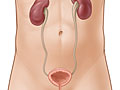
A kidney scan is a nuclear scanning test that is done to check kidney function or appearance.
During a scan to look at kidney function, a radioactive tracer substance is injected into a vein in the arm and then travels through the bloodstream to the kidneys. The tracer flows through the blood vessels in the kidneys and then is excreted into the urine. A special camera (gamma) takes pictures of the tracer in the kidneys. This helps show cell activity and function in the kidneys.
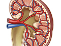
Many different kinds of kidney scans can be done. The types of kidney scans include a scan that looks at how blood flows to and through the kidneys, a scan that looks at the shape and size of the kidneys, and a scan that looks at how urine is made and flows out of the kidneys. Sometimes a doctor will do multiple scans at one time (for example, a triple renal study). Different radioactive tracers are used depending on what kind of scan is being done.
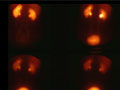
Areas of the kidneys where the tracer shows up in higher-than-normal amounts, such as in some types of cancer, result in bright or "hot" spots in the pictures. Areas where the tracer does not show up appear as dark or "cold" spots. Cold spots can mean narrowing or blockage of the blood vessels, pockets of fluid (cysts), some cancers, scarring, or pockets of infection (abscesses).
The amount of time it takes for the tracer to move through the kidney, collect in the urine, and drain into the bladder can also be seen in a kidney scan. This may be done to see how well the kidneys are working.
A kidney scan may be done instead of a kidney X-ray test called an intravenous pyelogram (IVP) for people who are allergic to the special dye (contrast material) used during the IVP.
Why It Is Done
A kidney scan is done to:
- Check the blood flow through the kidneys. Abnormal flow may mean narrowed renal arteries that can cause a type of high blood pressure called renovascular hypertension.
- See how a transplanted kidney is working.
- Check the extent of kidney damage caused by an injury or infection.
- Find an obstruction in the kidney or ureter, such as from a kidney stone.
- Find growths in the kidneys (rare).
How To Prepare
If you are breastfeeding, you may want to pump enough breast milk before the test to get through 1 to 2 days of feeding. The radioactive tracer used in this test can get into your breast milk and is not good for the baby.
How It Is Done
Before the test
You will need to remove any jewelry that might interfere with the scan. You may need to take off all or most of your clothes, depending on which area is being examined. (You may be allowed to keep on your underwear if it does not interfere with the test.) You will be given a cloth or paper covering to use during the test.
You may be asked to drink 2 to 3 glasses of water right before the scan.
During the test
The technologist cleans the site on your arm where the radioactive tracer will be injected. A small amount of the radioactive tracer is then injected. Medicine to increase your urine output (a diuretic) may also be injected. You may lie on your back on a table, stand, or sit upright. A large scanning camera will be positioned closely above your belly.
The camera will scan for radiation right after the radioactive tracer is injected. Scans may be taken every few minutes for about 30 minutes. More pictures may be taken 1 to 2 hours after the tracer was injected. The scans produce pictures as the tracer moves through your kidneys. You may also be given medicine to help the scans check for certain kidney functions.
A chart called a renogram may be made using the information from the kidney scan by plotting the movement of the tracer through the kidneys and recording it on a graph. A series of chart recordings is then made based on the amount of tracer uptake in the kidneys over a period of time. These recordings provide information about different phases of blood flow and kidney function.
You need to remain very still during each scan to avoid blurring the pictures. The camera does not produce any radiation, so you are not exposed to any more radiation while the scan is being done.
How long the test takes
A kidney scan usually takes about 30 minutes to 1 hour.
How It Feels
You may feel nothing at all from the needle puncture when the tracer is injected, or you may feel a brief sting or pinch as the needle goes through the skin. Otherwise, a kidney scan is usually painless. You may find it hard to remain still during the scan. Ask for a pillow or blanket to make yourself as comfortable as possible before the scan begins.
The test may be uncomfortable if you are having kidney pain. Try to relax by breathing slowly and deeply.
Risks
Allergic reactions to the radioactive tracer are rare.
Anytime you're exposed to radiation, there's a small chance of damage to cells or tissue. That's the case even with the low-level radioactive tracer used for this test. But the chance of damage is very low compared with the benefits of the test.
Steps you can take
Most of the tracer will leave your body through your urine or stool within a day. So be sure to flush the toilet right after you use it, and wash your hands well with soap and water. The amount of radiation in the tracer is very small. This means it isn't a risk for people to be around you after the test.
The radioactive tracer used in this test can get into your breast milk. Do not breastfeed your baby for 1 or 2 days after this test. During this time, you can give your baby breast milk you stored before the test, or you can give formula. Discard the breast milk you pump in the 1 or 2 days after the test.
You may have some soreness or swelling where the needle went in. These symptoms can usually be relieved by applying moist, warm compresses to your arm.
Results
The results of a kidney scan are usually available in 2 days.
Normal: | The radioactive tracer flows evenly to and through each kidney at the same time. The kidneys are working normally. |
|---|---|
The tracer flows from the kidneys into the urine, which then drains into the ureters and bladder. This process occurs within a normal time range. | |
The kidneys take up the radioactive tracer evenly. No "hot" spots or "cold" spots are seen. | |
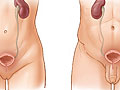
Abnormal: | The kidneys are not normal in shape, size, or location. |
The tracer does not flow evenly through the kidneys, meaning narrowing of, blockage of, or damage to the blood vessels or tissue in the kidneys. This may also mean poor kidney function. | |
The tracer collects in an area ("hot" spot) of a kidney. This might mean a tumor containing a higher-than-normal number of blood vessels. | |
An area of the kidney does not take up the tracer ("cold" spot). This may mean an abscess, cyst, or scarring. | |
The tracer does not pass from the kidneys into the urine and then through the ureters to the bladder. This can mean the movement of urine from the kidney is blocked. |
Credits
Current as of: July 26, 2023
Author: Healthwise Staff
Clinical Review Board
All Healthwise education is reviewed by a team that includes physicians, nurses, advanced practitioners, registered dieticians, and other healthcare professionals.
Current as of: July 26, 2023
Author: Healthwise Staff
Clinical Review Board
All Healthwise education is reviewed by a team that includes physicians, nurses, advanced practitioners, registered dieticians, and other healthcare professionals.
This information does not replace the advice of a doctor. Healthwise, Incorporated, disclaims any warranty or liability for your use of this information. Your use of this information means that you agree to the Terms of Use. Learn how we develop our content.